Photoacoustic imaging
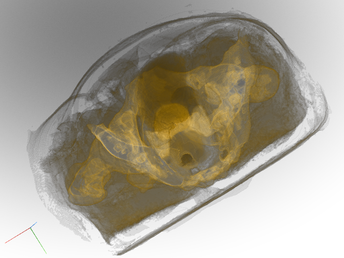
Realistic digital phantoms for prostate ultrasound and photoacoustic imaging
Yixuan Wu, Jacob Enders, Cheyenne Williams, Baichuan Jiang, James Wiskin, Michael B. Rothberg, Ayele H. Negussie, John Klock, Lindsey Hazen, Sheng Xu, Baris Turkbey, Peter A. Pinto, Bradford J. Wood, Emad M. Boctor
SPIE Medical Imaging, 2024
Data | PDF
We introduce the first realistic digital phantoms for prostate ultrasound and photoacoustic (PA) imaging in the male pelvic region. Each digital phantom set contains five parameters: speed of sound, density, acoustic attenuation, optical absorption, and reduced optical scattering.
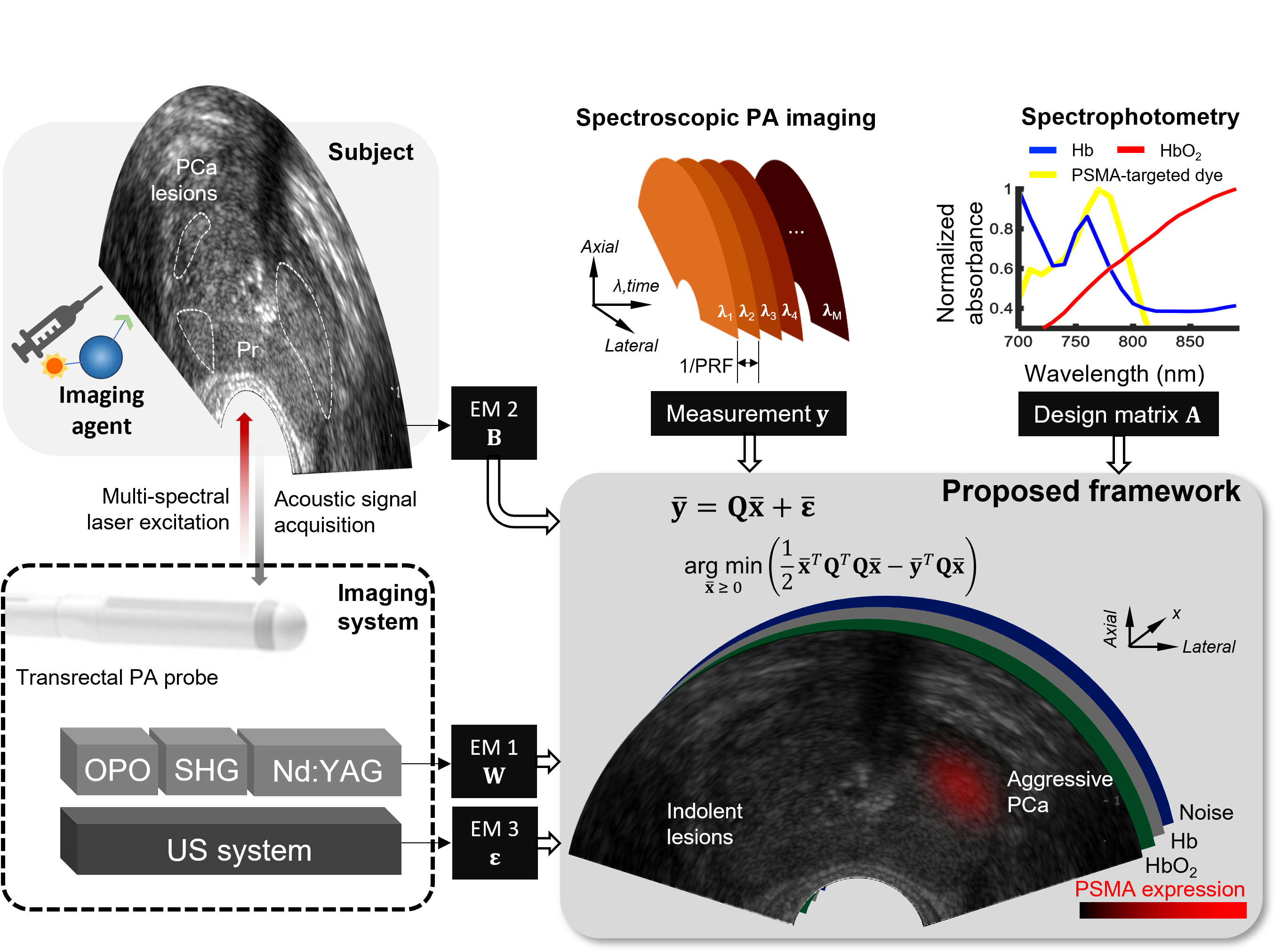
System-level optimization in spectroscopic photoacoustic imaging of prostate cancer
Yixuan Wu*, Jeeun Kang*, Wojciech G.Lesniak, Ala Lisok, Haichong K.Zhang, Russell H.Taylor, Martin G.Pomper, Emad M.Boctor
Photoacoustics, 2022
PDF
One challenge for prostate cancer detection is to have both noninvasive and high-contrast imaging of deep prostate tissues. Photoacoustic (PA) imaging has been shown to be able to cater to this unmet need, meanwhile providing functional and quantitative information. This study presents a system-level optimization of spectroscopic PA imaging for prostate cancer detection in three folds: system noise denoising, wavelength selection, and frame averaging. The proposed framework is validated both in simulation and in vivo and showed the capability for more sensitive and faster prostate cancer detection.
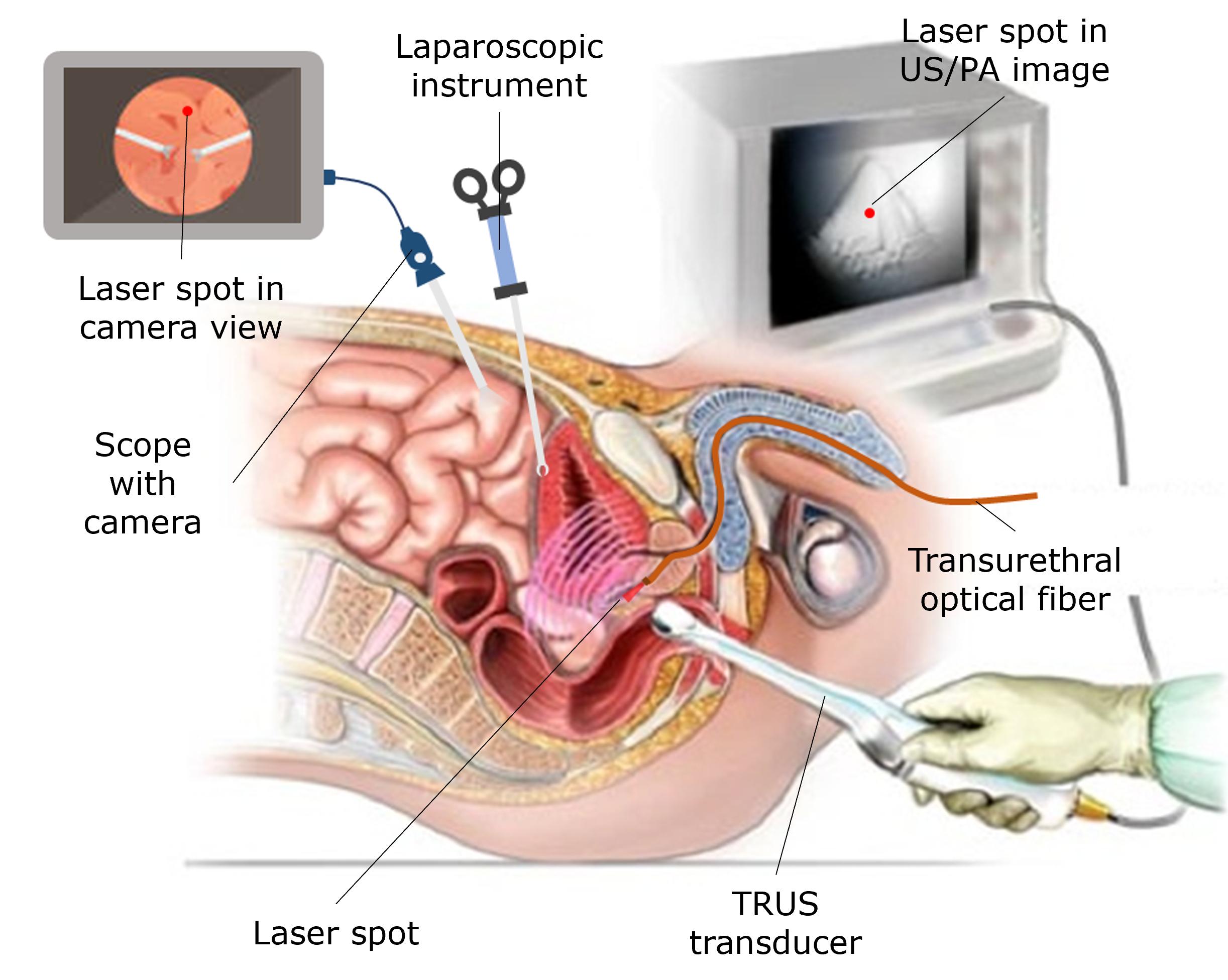
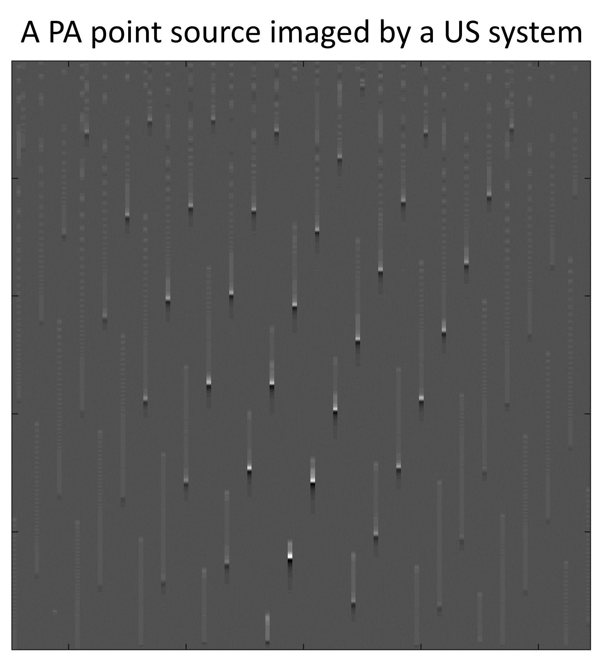
Feasibility of using low-energy pulsed laser diode on clinical ultrasound platforms for photoacoustic and transrectal ultrasound guided laparoscopic prostatectomy
Yixuan Wu, Baichuan Jiang, Hyunwoo Song, Keshuai Xu, Hamid Moradi, Emad M Boctor
IEEE International Ultrasonics Symposium (IUS), 2022
Video | PDF
Here we present a feasibility study of adding a low-cost, low-energy, and compact pulsed laser diode (PLD) system on clinical ultrasound (US) platforms for photoacoustic (PA) point marker imaging. The size of the PLD plus a driver is no larger than a hand-held US probe, and the energy per pulse of the PLD is in µJ level, which is 3 to 4 orders of magnitude lower than conventional PA lasers. We tackled two major challenges in this work, one is the very low energy per pulse of the PLD, and the other is the inaccessible beamformer on clinical US machines.
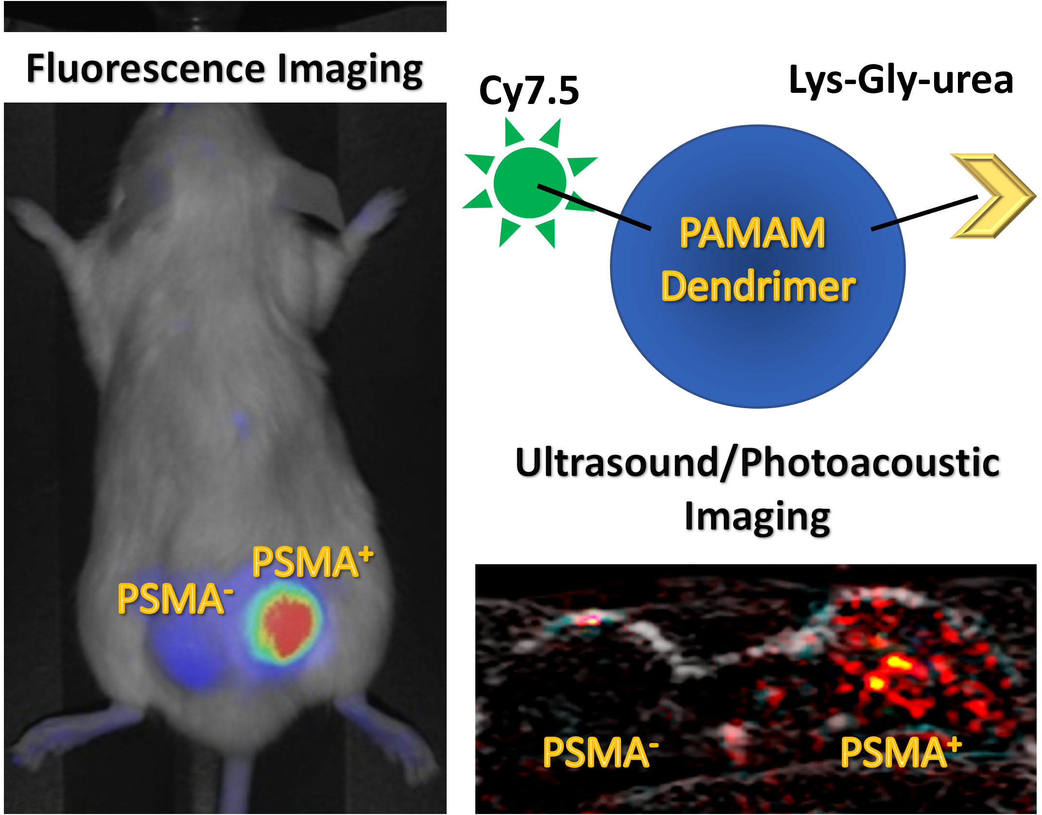
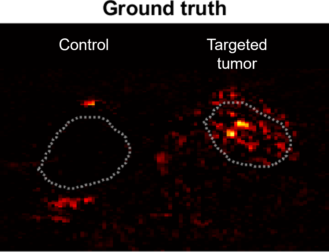
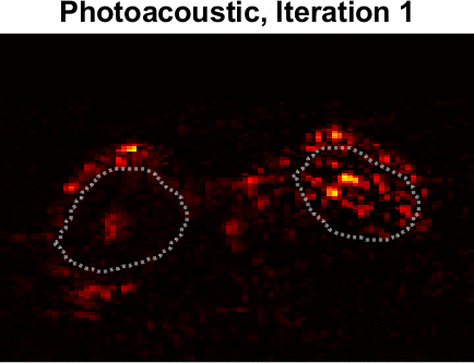
Dual contrast agents for fluorescence and photoacoustic imaging: evaluation in a murine model of prostate cancer
Wojciech G. Lesniak*, Yixuan Wu*, Jeeun Kang, Srikanth Boinapally, Sangeeta Ray Banerjee, Ala Lisok, Anna Jablonska, Emad M. Boctor, Martin G. Pomper
Nanoscale, 2021; IEEE International Ultrasonics Symposium (IUS), 2020
Video | PDF | Patent
Prostate-specific membrane antigen (PSMA) is a promising diagnostic and therapeutic target for prostate cancer (PC). Poly(amidoamine) [PAMAM] dendrimers serve as versatile scaffolds for imaging agents and drug delivery that can be tailored to different sizes and compositions depending upon the application. In this work, we developed PSMA-targeted Poly(amidoamine) [PAMAM] dendrimers for real-time detection of PC using fluorescence (FL) and photoacoustic (PA) imaging. Such agents may prove useful in PC cancer detection and subsequent surgical guidance during excision of PSMA-expressing lesions.
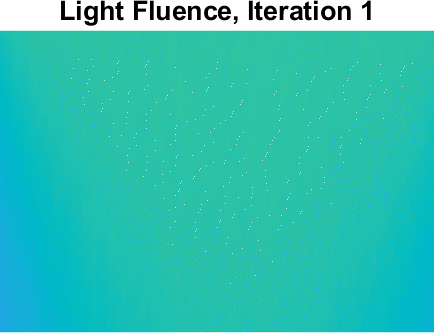
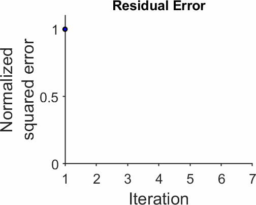
Iterative fluence compensation and spectral unmixing for spectroscopic photoacoustic imaging
Yixuan Wu†, Jeeun Kang, Wojciech G. Lesniak, Martin G. Pomper, Emad M. Boctor
IEEE International Ultrasonics Symposium (IUS), 2021
Video | PDF
An iterative fluence compensation and spectral unmixing algorithm for spectroscopic photoacoustic imaging (SPA) is described. The algorithm focuses on solving the optical inverse problem. It employs optical prior knowledge of tissues, leverages a Monte Carlo simulator for fluence estimation, and assumes a linear mixed model for absorption, scattering, and anisotropy. After an initial guess of the tissue composition, the algorithm sequentially estimates the light fluence, solves for tissue concentrations in spectral unmixing, and updates the optical parameters iteratively until the estimated initial pressure converges to the measurement.
An economic photoacoustic imaging platform using automatic laser synchronization and inverse beamforming
Yixuan Wu, Haichong K.Zhang, Jeeun Kang, Emad M.Boctor
Ultrasonics, 2020
PDF
We present a proof-of-concept of an automatic integration of photoacoustic (PA) imaging on clinical ultrasound (US) imaging platforms. Here we tackle two critical challenges: the laser synchronization and the inaccessibility to the beamformer core embedded in commercial US imaging platform. In particular, the line trigger frequency (LTF) estimation and the asynchronous synthetic aperture inverse beamforming (ASAIB) were developed and evaluated in both k-Wave simulation and phantom experiment. The proposed method is an economical solution to enable PA imaging on a greater number of US equipment to further thrive the PA imaging research community.
Ultrasound tomography
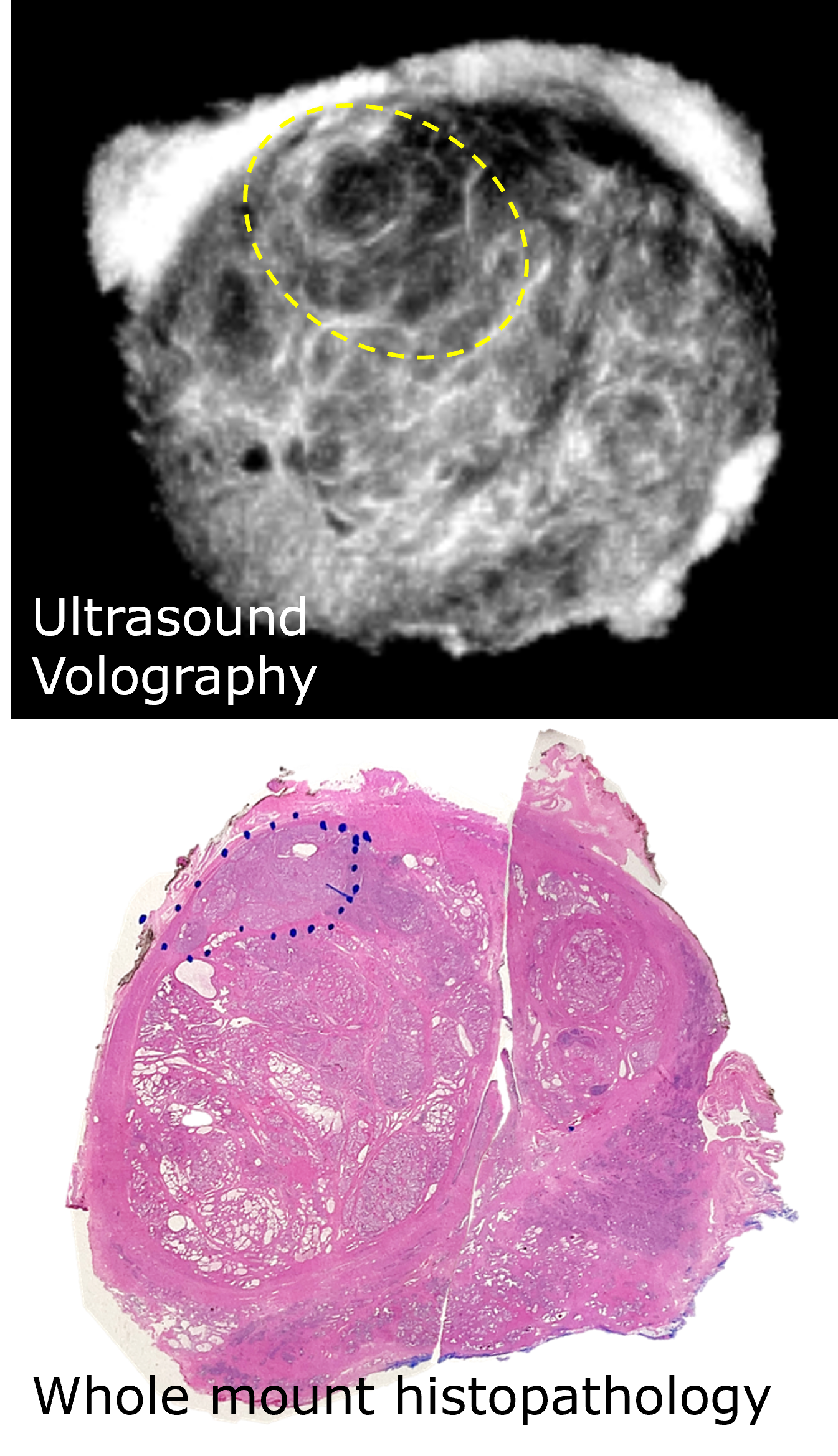
Ultrasound tomography for prostate cancer imaging: An ex vivo preliminary study
Jacob J. Enders, Cheyenne Williams, Michael B. Rothberg, Zoe Blake, Jibriel Noun, Yixuan Wu, James Wiskin, Michael Daneshvar, Reza Seifabadi, Ayele H. Negussie, Peter L. Choyke, Emad M. Boctor, Antoun Toubaji, Maria J. Merino, Baris Turkbey, Bradford J. Wood, Peter A. Pinto
Annual Meeting of Engineering and Urology Society (EUS), 2022
Best Abstract Award
Imaging of prostate cancer with 3D ultrasound tomography
James W Wiskin, Jacob Enders, Ismail Turkbey, Michael Rothberg, Maria Merino, Sheng Xu, Emad Boctor, Yixuan Wu, Ayele Negussie, John Klock, Brad Wood, Peter Pinto
SPIE Medical Imaging, 2022
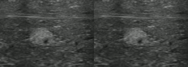
Prostate ultrasound tomography (UT): correlation with MRI and whole mount histopathology
Cheyenne Williams, Michael Daneshvar, Yixuan Wu, Jeunice Owens-Walton, Nitin Yerram, Patrick T. Gomella, Luke P. OConnor, Nabila Khondakar, Michael Ahdoot, Ayele Negussie, Sheng Xu, Baris Turkbey, Maria Merino, Emad Boctor, James Wiskin, Bradford Wood, Peter Pinto
American Urological Association (AUA) Annual Meeting, 2021
Video | PDF (EUS) | Link (AUA)
With recent developments, state-of-the-art ultrasound tomography (UT) is now able to provide submillimeter-resolution imaging. Compared to conventional pulse-echo ultrasound (US) imaging, UT provides quantitative US transmission that characterizes speed of sound and constructs speckle-free, refraction-corrected 360-degree-compounded reflection images. Given the success of UT for accurately diagnosing breast cancers in patterns consistent with MRI, we sought to define feasibility of UT for prostate cancer imaging.
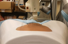
Dual-robotic ultrasound system for in vivo prostate tomography
Kevin M Gilboy, Yixuan Wu, Bradford J Wood, Emad M Boctor, Russell H Taylor
Advances in Simplifying Medical UltraSound (ASMUS) MICCAI Workshop, 2020
Best Paper Runner-up
PDF
Ultrasound tomography (UST) offers quantitative anatomical tissue characterization. While most existing UST systems focused on submerging target anatomy in a transducer-lined cylindrical water tank, they are not practical for deep anatomy such as prostate. This work outlines and validates a clinical workflow and real-time motion framework for a novel dual-robotic approach to in vivo prostate imaging: one arm wielding a linear abdominal probe, the other wielding a linear transrectal ultrasound probe.
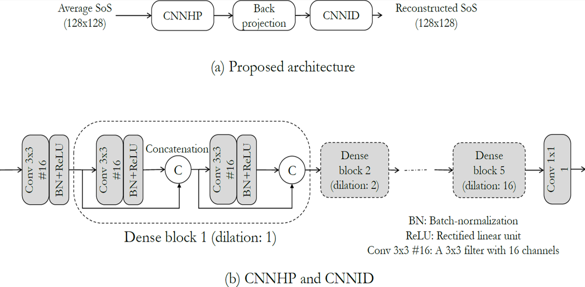
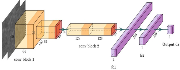
CNN and back-projection: limited angle ultrasound tomography for speed of sound estimation
Emran Mohammad Abu Anas, Alexis Cheng, Reza Seifabadi, Yixuan Wu, Fereshteh Aalamifar, Bradford Wood, Arman Rahmim, Emad M Boctor
SPIE Medical Imaging, 2019
Video | PDF
Ultrasound tomography has the potential to quantify tissue acoustic properties for advanced clinical diagnosis. However, the location of most human anatomies limits the tomography to a few angles that leads the reconstruction a challenging problem. In this work, a deep convolutional neural networks-based technique is presented to estimate the speed of sound of tissue from limited-angle data.
Others
| Without tracking | With tracking |
Stabilized ultrasound imaging of a moving object using 2D B-mode images and convolutional neural network
Tian Xie, Mahya Shahbazi, Yixuan Wu, Russell H. Taylor, Emad M. Boctor
SPIE Medical Imaging, 2020
Video | PDF
We present a co-robotic ultrasound imaging system that tracks lesions undergoing physiological motions, e.g., breathing, using 2D B-mode images. The approach embeds the in-plane and out-of-plane transformation estimation in a proportional joint velocity controller to minimize the 6-degree-of-freedom (DoF) transformation error. Specifically, we propose a new method to estimate the out-of-plane translation using a convolutional neural network based on speckle decorrelation. The network is trained on anatomically featureless gray-scale B-mode images and is generalized to different tissue phantoms. The tracking algorithm is validated in simulation with mimicked respiratory motions, which demonstrates the feasibility of stabilizing biopsy through ultrasound guidance.
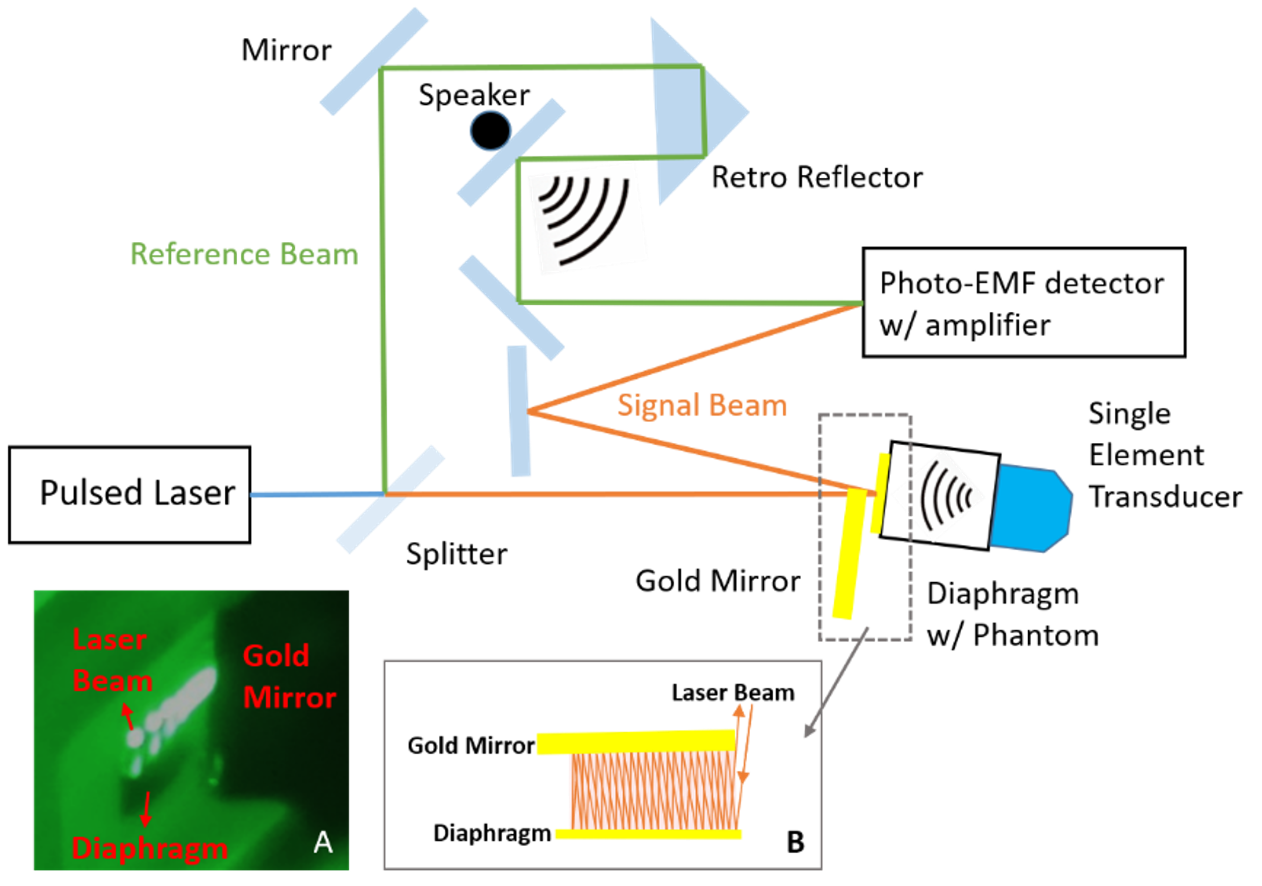
Ultrasound signal detection with multi-bounce laser microphone
Qianqian Wan, ChenChia Wang, Keshuai Xu, Jeeun Kang, Yixuan Wu, Sudhir B Trivedi, Peter Gehlbach, Emad Boctor
IEEE International Ultrasonics Symposium (IUS), 2020
Link
The multi-bounce laser microphone utilizes optical methods to detect the displacement of a gold-covered thin film diaphragm caused by ultrasound signal pressure waves. This sensitive technique provides new opportunities for advanced ultrasound imaging in terms of a higher detection signal-to-noise ratio (SNR) in a broader spectrum than conventional ultrasonic transducers. The system was previously developed for detecting acoustic signatures generated by explosives and were limited to lower than 10 kHz in frequency. In this work we demonstrate its feasibility for biomedical imaging applications.